Introduction
The NHS defines bladder weakness, also known as urinary incontinence, as “the unintentional passing
of urine”.
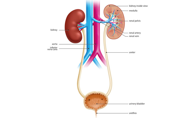
To understand why this can occur, it can help to have an understanding of the body’s urinary system, which is composed of various interconnected structures that make, store and excrete urine – one of the body’s waste products.
- Sphincter: this ring of muscle surrounds the opening of the bladder to keep urine inside and prevent leaks
- Pelvic floor muscles: these surround and support the bladder and the urethra. The stronger they are, the better they are able to prevent leaks
- Prostate gland: located in men between the penis and the bladder, the prostate gland is involved in semen production. If it becomes enlarged and presses on the bladder and urethra, it can lead to bladder weakness
- Kidneys: two bean-shaped organs, each about the size of a fist, located near the middle of the back, just below the ribcage. Their job is to remove waste and excess fluid from the blood and produce urine
- Ureters: each kidney is connected to the bladder by a thin tube called a ureter. Muscles in the ureter walls constantly contract and relax to take urine from the kidneys to the bladder
- Bladder: this muscular bag stores urine, stretching and expanding as it fills up. Nerve signals tell the brain that the bladder needs to be emptied. Although it varies, the average person empties their bladder four to eight times a day and sometimes during the night
- Urethra: this tube takes urine from the bladder to outside of the body. It is relatively short in women, and longer in men.

